Applications

Intended Use

The device is designed to measure, by applying the technique of EIT, the regional changes in thoracic impedance in the measured area due to changes in content of gas and liquid (ventilation or perfusion) and short-term changes in end-expiratory lung volume (EELV).

Multiple Departments
  • Intensive Care Unit/Operating Room/Emergency Department

  • Department of Respiratory

  • Department of Rehabilitation

  • Department of Cardiothoracic Surgery

  • Other departments requiring ventilation/perfusion monitoring

Multiple Diseases
  • Pneumonia

  • ARDS

  • COPD

  • Pleural effusion

  • Hemothorax

  • Pneumothorax

  • Pulmonary embolism

  • Pulmonary arterial hypertension

Operation Guide
  • Switching on the EIT System

  • Device Check

  • Connecting the Electrode belt, Patient cable, and Equipment cable Preparing the patient

  • Check Signal Quality

  • Enter Patient Information on the Start Page

  • Set Cut Off Frequency

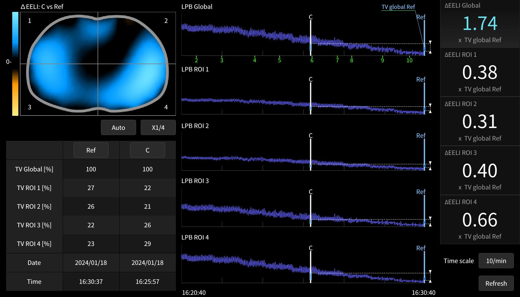
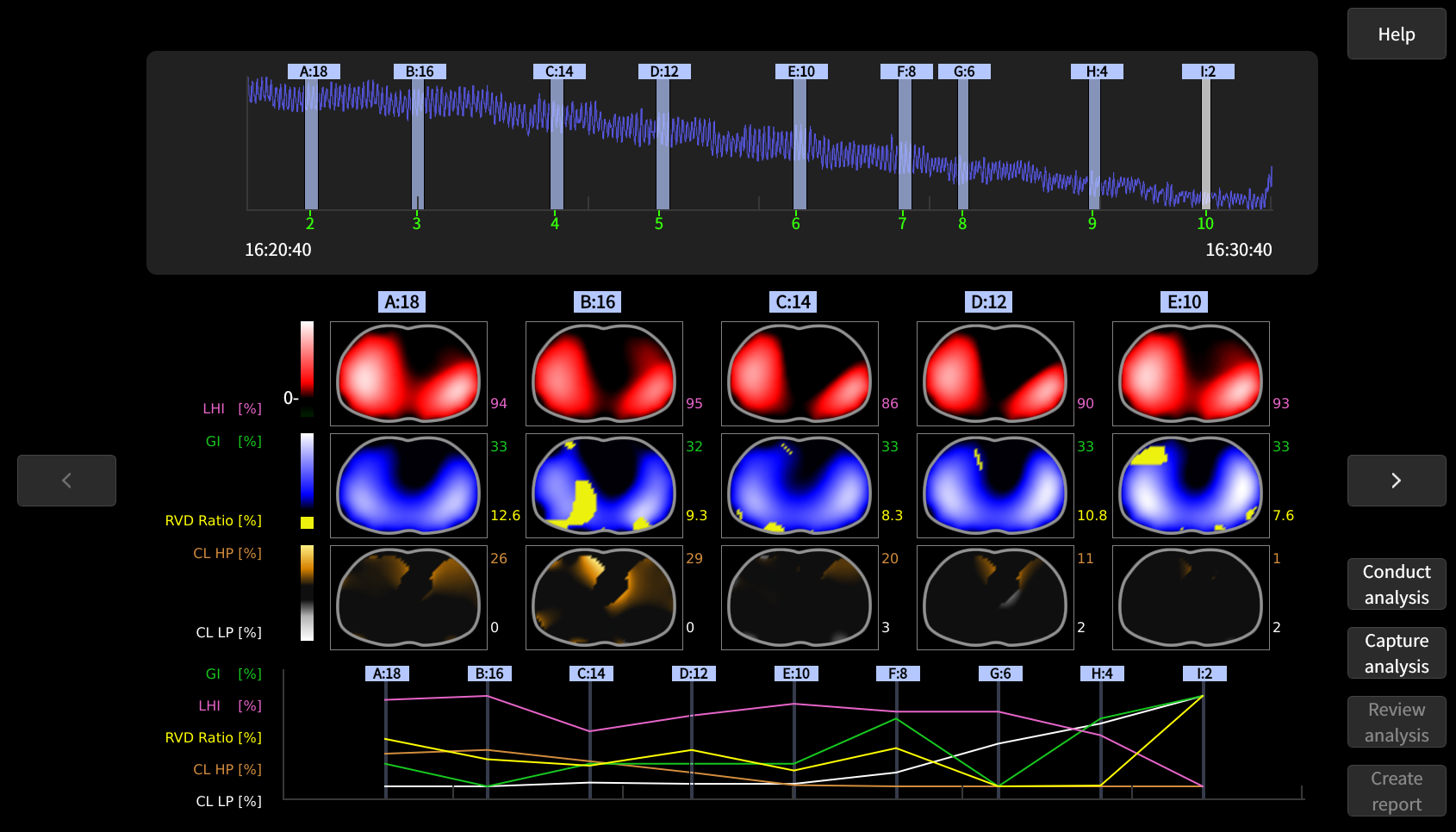
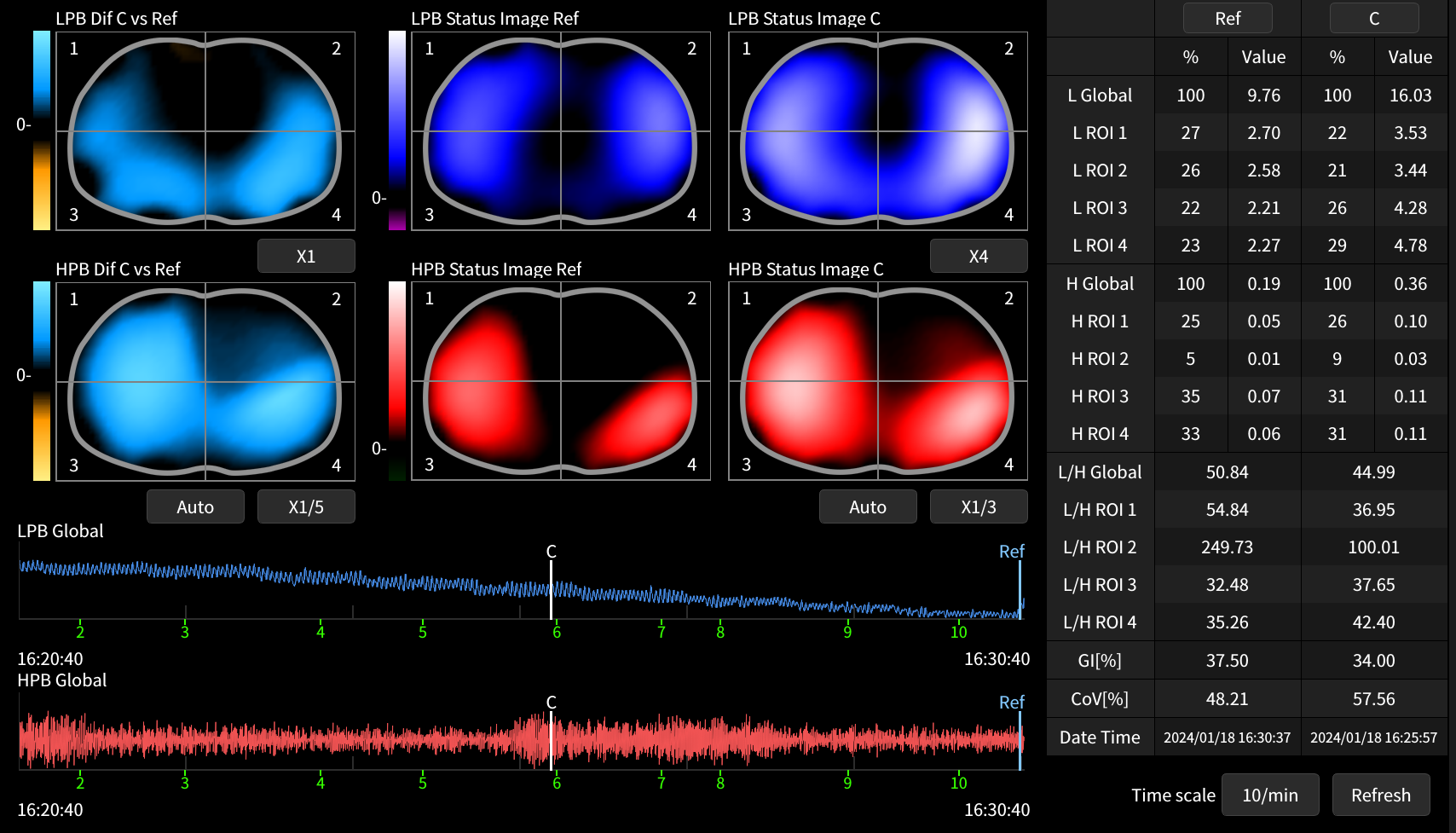
  • Start Monitoring and Assessing Using Main, ΔEELI, Trends, Analysis and Match Views

  • Disinfection


Highlights

Warnings

For your safety and that of your patients, please operate the device in strict accordance with the IFU.

The device is not intended for home use and must be operated by skilled/trained clinical professionals.

Monitoring information is provided for reference purpose only and should not be used as the sole basis for treatment or diagnosis.

The installation, operation, expansion, change, modification, and maintenance of the device must be carried out only by technicians authorized by Infivision.

Features
Exclusive Technology
The world’s pioneer in pulse wave perfusion imaging, which is contrast agent free and completely non-invasive.
Real Time
To see the pulmonary functional changes without any delay.
Non-Invasive
Ensuring safety, minimizing risks and enhancing patient comfort.
Radiation Free
Eliminates the risks associated with radiation, ensuring patient well-being with repeated use.
Functional Imaging
Functional imaging assists health workers in observing changes in medical conditions by visualizing pulmonary ventilation and perfusion alterations.
Bedside
Safeguard stands next to the patients, saving both cost & time.
Portable Design
Ultra-portable, easy to carry, ideal for on-the- spot ICU use.
Facilitating Research
Supports data export and empowers in-depth scientific research by providing specialized data analysis software (ETSA), enabling comprehensive study and innovation in medical science.
Diverse analysis tools
Main View ( Main Veiw & Large Mode )

Provides several sections to help quickly assess key pulmonary function information which include:

  • Continuous, dynamic images of ventilation and perfusion

  • Status images of overall and region-of-interest distribution of ventilation and perfusion

  • Display of numerical values or percentages for regional respiratory and perfusion impedance changes & display ratio of center of ventilation COV%.

ΔEELI View

This view is used to assess the changes of End Expiratory Lung Impedance (ΔEELI) to,

  • Evaluate changes in ΔEELV which reflects alterations in Functional Residual Capacity (FRC) -(Ex - After modifying the Positive End-Expiratory Pressure (PEEP), applying recruitment maneuvers, or proning the patient)

  • Aiding in the detection of possible recruitment of individual lung areas

  • Aiding in the detection of possible de-recruitment of individual lung areas

Analysis View

Allows the analysis of regional compliance changes and delays in regional ventilation in addition to the evaluation of ventilation & perfusion distribution.

  • Automatically evaluate progressive and regressive PEEP adjustments to identify the optimal PEEP that effectively balances between over-expansion and collapse.

  • Evaluate the potential for lung recruitment.

  • Shows the correlation degree between ventilation and perfusion, based on impedance variation status images.

Match View

Facilitating research by giving more information such as MI (match index), DI (deadspace index), SI (shunt index) and related waveforms to observe and explore more.

Trend View

Compares the respiratory and perfusion images corresponding to any two giventime points to

  • Continuous, dynamic images of ventilation and perfusion

  • Status images of overall and region-of-interest distribution of ventilation and perfusion

  • Display of numerical values or percentages for regional respiratory and perfusion impedance changes & display ratio of center of ventilation COV%.

How may we assist you?