Applications
1.Quick assessment of diagnosis and treatment
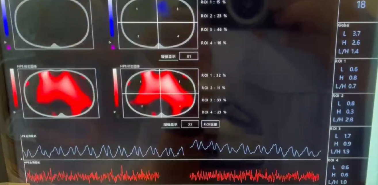
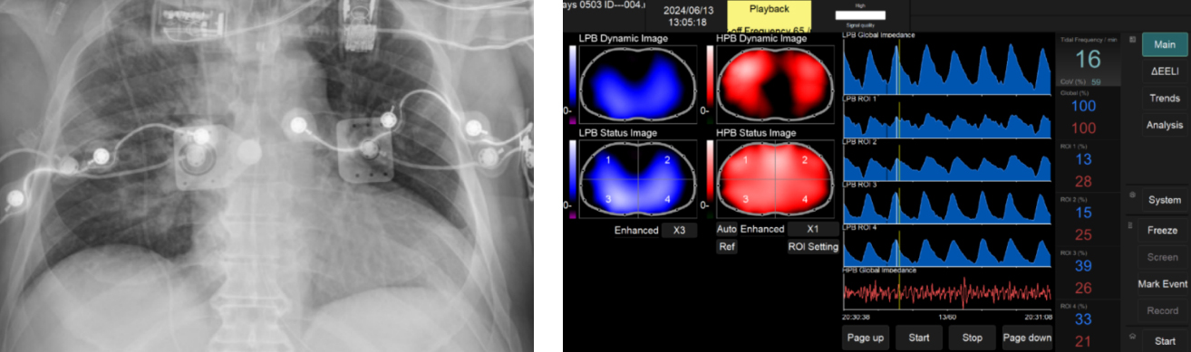
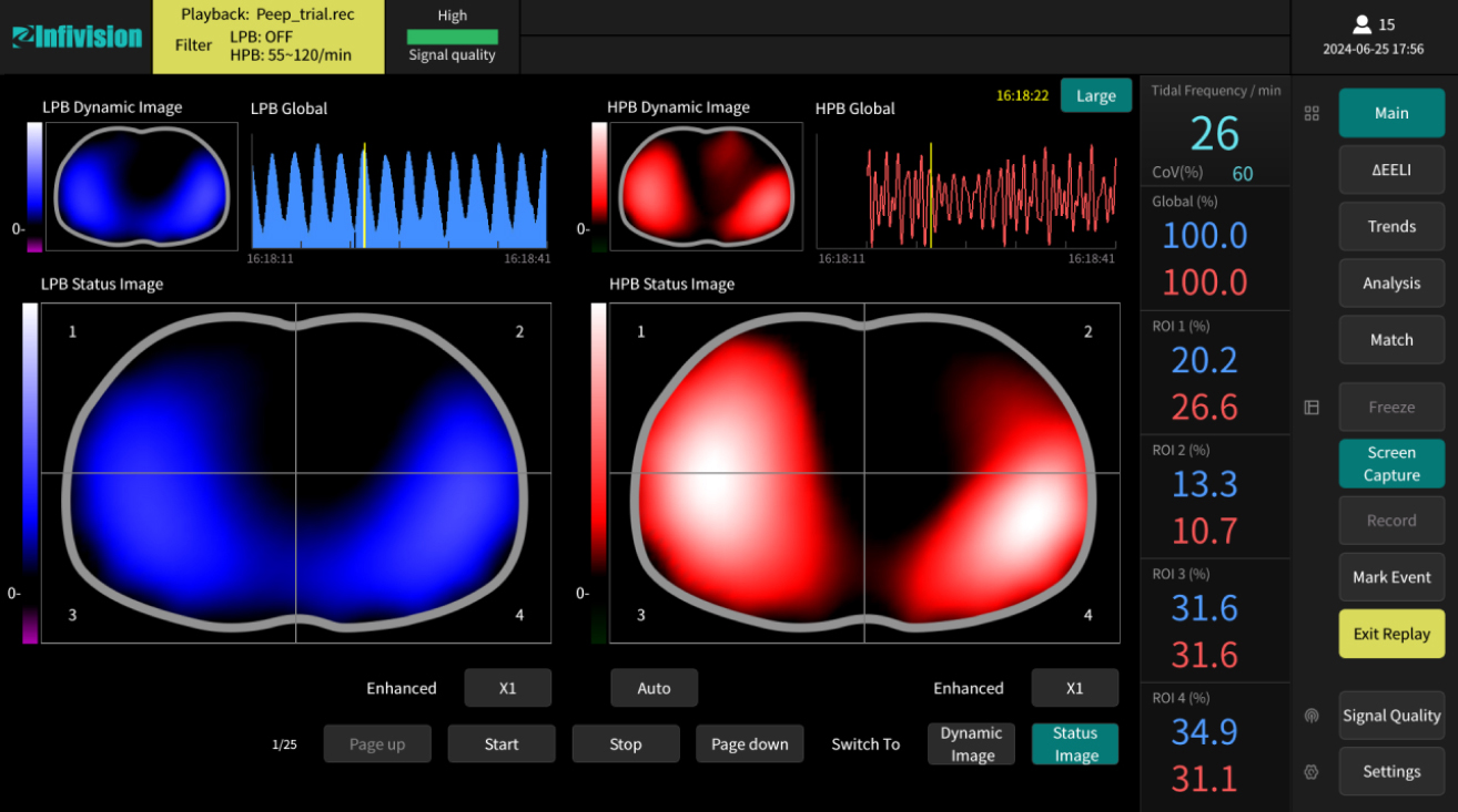
Case 1 (lCU) Exclusion of PE Diagnosis
Exclusion of pulmonary embolism diagnosis in a patient with right ventricular dysfunction, cardiogenic shock, kidney insufficiency, and ARDS with right-sided pneumonia. These conditions were confirmed in the trauma room using the Infivision ET1000.
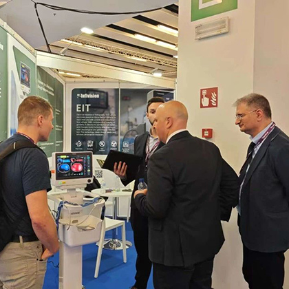
EIT Infivision in trauma room.
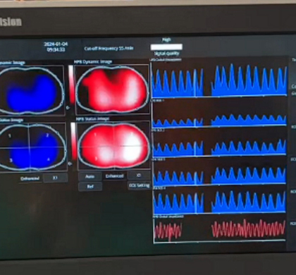
Right-sided pneumonia with bilateral perfusion.
Right ventricular enlargement was confirmed with echocardiogram.
Courtesy of Dr. Baldassare Ferro, MD., Ospedali Riuniti di Livorno, Italia
Case 2 (OR) Acute low SpO2 after lower limb trauma surgery
Patient A, male, 41 years old. The patient underwent emergency lower limb trauma surgery on July 22nd. He was weaned and extubated on July 23rd at 11:33 AM. At 2:48 PM, the respiratory rate increased to 37 breaths per minute, and SpO2 began to decline. Differential diagnosis: 1. Acute airway obstruction? 2. ARDS? 3. PE?
When we used the bedside Infivision ET1000 to visualize pulmonary ventilation and perfusion in real-time, we observed that the ventilation signal was almost absent, while the perfusion signal was still partially present.
Reintubation was performed. Fiberoptic bronchoscopy therapy was administered to the patient after utilizing the Infivision ET1000 to locate the specific area of abnormal ventilation. A large to moderate amount of secretions was observed through fiberoptic bronchoscopy in both the right and left main bronchi.
SpO2 returned to 97% shortly after suctioning.
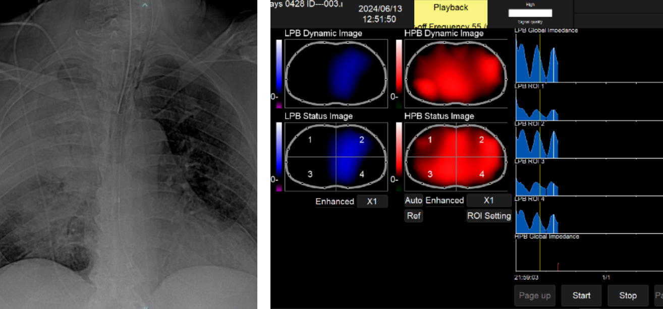
2. Continuously monitoring
Septic shock and kidney dysfunction and severe ARDS
Day 1
A male patient in the ICU with septic shock, kidney dysfunction, and right lung ARDS.
Courtesy of Dr. Baldassare Ferro, MD., Ospedali Riuniti di Livorno, Italia
Day 2
X-ray and Infivision ET1000 showed worsening of ARDS.
Patient with ARDS, severe respiratory failure (PaO2/FiO2=60mmHg), not candidate to ECMO and prone position due to hemodynamic instability. Inhomogeneous involvement and near complete atelectasis of right lung not recruited by protective ventilation in the presence of equal perfusion (Infivision ET1000).
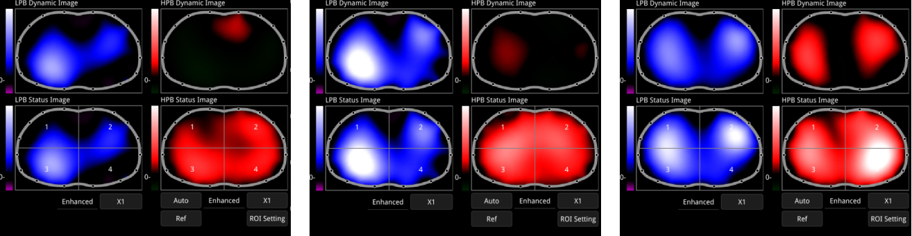
Day 4
X-ray and Infivision ET1000 showed right lung was getting better through independent lung ventilation with double lumen intubation guided by Infivision ET1000 applying 20cmH2O of PEEP on right lung and protective ventilation on left determined slow recruitment of closed lung ameliorating oxygenation, and conventional imaging.
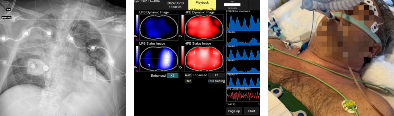
Courtesy of Dr. Baldassarre Ferro, MD., Ospedali Riuniti di Livorno, Italia
Day 6
X-ray and Infivision ET1000 showed significant improvement after therapy: both ventilation and perfusion were improving, with ventilation becoming more homogeneous.
Courtesy of Dr. Baldassarre Ferro, MD., Ospedali Riuniti di Livorno, Italia
Day 7
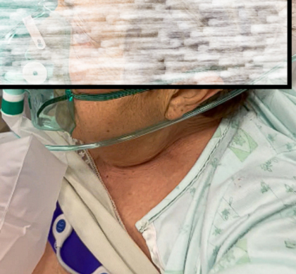
The skin shows no marks after wearing the belt for seven days.
3. Individualized respiratory management
Individualized PEEP Titration
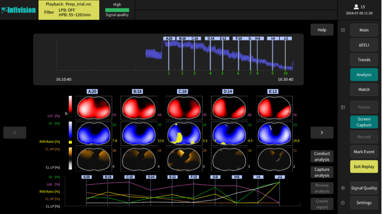
Prone Positioning
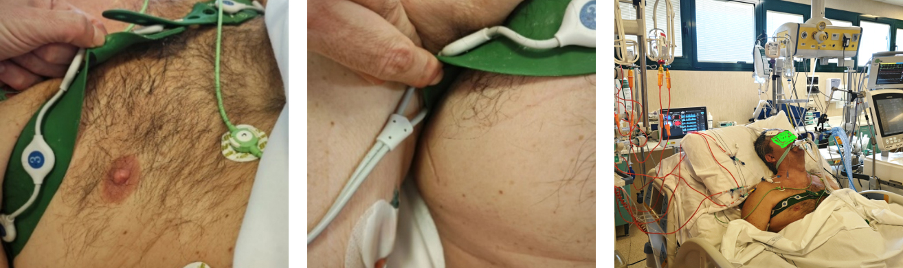
4.Ease of use on bedside

Easy and intuitive patient observation.
Change batch
Copyright © 2022 Infivision Medical lmaging Technology Co.,Ltd Beijing ICP 2022020544-2 (Beijing) network medical equipment information (2023) No. 00060
Beijing public network security 11011502006084 webmaster statistics